
Aroid Leaf Disease: Diagnosing the Silent Killers
Aroid leaf disease forensics: how to tell bacterial blight (Xanthomonas) from fungal spots and abiotic damage by lesion shape, halo, and the ooze test, and why sanitation, quarantine, and a fan beat any spray.
Samuel Reed · Published 2026-01-12 · 27 min read
Key Takeaways
- Keep foliage dry when a pathogen may be present. A wet leaf surface gives bacteria such as Xanthomonas a film for movement, so air movement and drier leaves lower risk. Guttation droplets are a possible entry point, not proof of infection.
- Lesion shape, halo, texture, and location are clues rather than a diagnosis. Greasy V-shaped lesions with bright halos can lean bacterial, dry concentric rings can lean fungal, and crispy tips after feeding can lean abiotic. Chilling, water molds, and mixed infections overlap heavily.
- Confirmed systemic blight in Anthurium has a poor prognosis. There is no reliable home cure, and copper and antibiotics have practical limits and legal label restrictions. Isolate and, where appropriate, dispose of confirmed infected plants rather than relying on chemistry.
- Quarantine reduces risk without proving a plant is clean. New plants should be watched for at least 30 days, but Xanthomonas can remain symptom-free for months. Clean stock and testing matter more than any single waiting period.
- Tool sanitation limits cross-contamination. Follow the disinfectant label for dilution and contact time, work in a ventilated area, and never mix bleach and alcohol.
Aroid leaf spots rarely identify their pathogen by appearance alone. Use lesion shape, texture, spread, plant history, and a few safe home observations to decide what to isolate and document, then confirm serious or spreading cases through a diagnostic lab before treating or discarding a plant.
The Physiology Behind Aroid Leaf Disease Vulnerability
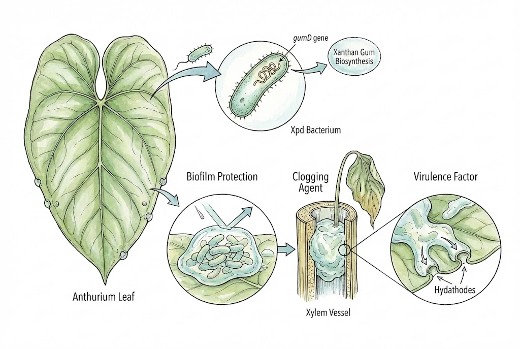
Start with what you can observe. Water at the leaf edge, wounds, wet foliage, and shared tools can all give disease an opportunity to spread.
The Hydathode (A Vulnerable Entry Point)
Most people are familiar with stomata (the microscopic pores on the underside of leaves used for gas exchange).
However, Aroids also possess specialized pores called hydathodes, located along the leaf margins and sometimes at the leaf tip.
Hydathodes are pores at leaf edges and tips. Under very wet conditions, a plant can push droplets from them, called guttation.
Guttation or condensation alone does not diagnose disease. Wipe up standing droplets, avoid splashing leaves, and improve airflow if foliage stays wet.
Hydathode entry is one reason a V-shaped, margin-in lesion is a recognised pattern for this disease. Infection near the pore can progress inward toward a vein.
It is not the only way lesions form, and stomatal entry can produce more circular spots.
Seeing guttation droplets by itself does not mean your plant is infected or at high risk. Risk depends on whether a pathogen is actually present, plus splashing water and overall plant health.
Bacterial blight of anthurium and other aroids (UF/IFAS)
The Cuticle and the Cell Wall
The first line of defense for any leaf is the cuticle. A waxy, hydrophobic layer that prevents water loss and creates a barrier against pathogens.
Leaf surfaces vary widely across aroids. Many Philodendrons feel thick and waxy, while Anthuriums such as Anthurium warocqueanum have thinner, velvety leaves.
It is tempting to say this single difference is why Anthurium is so often affected by Xanthomonas phaseoli pv. dieffenbachiae (Xpd), but the evidence does not support a clean cuticle-thickness ranking.
Susceptibility differs by cultivar and bacterial strain, and formal records describe both leaf and systemic infection in Anthurium. Surface texture alone does not predict how vulnerable a plant is.
Once bacteria are inside a leaf, they meet the cell wall, which is built largely of cellulose and pectin (carbohydrates that hold cells together).
Bacteria such as Xanthomonas and Pectobacterium (a soft-rot organism) can produce enzymes, pectinases and cellulases, that break down these bonds, though the two groups differ in how strongly and in what role they do so.
A water-soaked spot can reflect this kind of enzymatic breakdown, but it is not proof of it. Chilling injury, water molds, and simple mechanical damage can all look water-soaked too, so the appearance alone does not confirm a bacterial cause.
Where maceration does occur, cell walls collapse, cytoplasm leaks out, and the air spaces that normally make a leaf look opaque fill with liquid.
That changes how light passes through the tissue, which is why such a spot can look translucent or greasy against a strong light (a useful clue, not a pathogen-specific marker).
The Vascular System
When Xpd becomes systemic, it reaches the xylem vessels that carry water from the roots to the leaves.
Not every leaf infection progresses this far, so systemic spread is possible rather than inevitable.
Bacteria that reach the vascular system can multiply and produce extracellular polysaccharides (slime).
This can turn a localised leaf problem into a whole-plant one, with bacteria travelling down the petiole, into the stem, and into other leaves. A course documented especially in Anthurium.
Vascular blockage is one explanation for why a plant can wilt while its medium is still wet.
But wet-soil wilt has other common causes (root rot, waterlogging, other vascular pathogens such as Ralstonia), so a wilting plant with brown vascular streaks needs a lab test to confirm what is actually responsible rather than an assumption of Xpd.
The Primary Pathogen in Aroid Leaf Disease – Xanthomonas
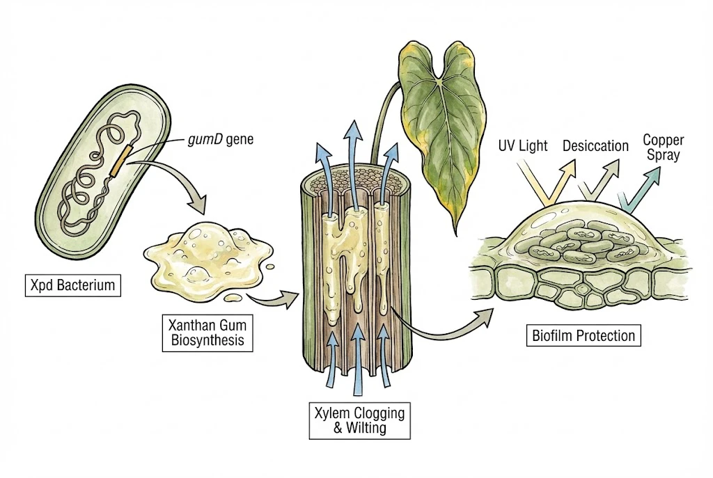
Xanthomonas phaseoli pv. dieffenbachiae (Xpd) is a well-documented and serious bacterial pathogen of Anthurium and related aroids.
Once bacteria are inside a leaf, surface sprays cannot reliably clear the infection. That is why early isolation, dry foliage, sanitation, and laboratory confirmation matter more than chasing a home cure.
The Latency Period (Asymptomatic Incubation)
One of the hardest things about Xpd is that an infected plant can look healthy.
Extension sources note that a plant can carry Xpd and stay symptom-free for months while bacteria are present and possibly multiplying. This is why clean, screened stock matters, especially with tissue-cultured plantlets that were not rigorously tested.
A plausible scenario. You acquire an Anthurium that looks perfect, even from a well-regarded grower, and add it to your collection.
Weeks later, after a heat spell or irregular watering, it begins to show symptoms.
It is often assumed that stress and warm temperatures around 28–32°C wake up dormant bacteria, but that trigger story is not established for Xpd. A symptom-free infection is not the same as a dormant one. Incubation figures also come from specific study conditions. One nested-PCR study reported latency around 16–28 days, so treat a 14–30 day range as condition-dependent rather than a fixed rule.
Whatever the exact timing, the practical point holds. Because an infected carrier can look fine for a long time, it may spread bacteria to other plants through shared water, tools, or contact before it ever shows symptoms.
Diagnosing the Progression (A Common Sequence)
Xpd often moves through the rough sequence below, and knowing it helps you read symptoms. Real cases do not always follow every step in order, so use these as recognisable phases, not a fixed timetable.
Stage 1. The Water-Soaked Lesion (Initial Infection)
An early sign is subtle and often appears on the underside of the leaf, near the margins (where the hydathodes are).
You may see small, angular, dark-green, greasy or water-soaked spots. Some early lesions are more star-shaped or, with stomatal entry, more circular.
Hold the leaf up to a strong light. A water-soaked bacterial lesion often looks translucent, like stained glass, because light passes through liquid-filled spaces. This is a helpful clue rather than a confirmation. Chilling injury and water molds can also look water-soaked, and a dry Xpd lesion can look dark rather than translucent, so opacity does not prove a fungal or abiotic cause.
Early lesions are usually small, often a few millimetres, and can expand as bacteria multiply and break down mesophyll tissue.
Stage 2. The Chlorotic (Yellow) Halo
As the colony expands, a bright lemon-yellow band often forms between the dead tissue and the healthy green leaf.
A distinct, intense halo that runs ahead of the necrosis is a useful pointer toward a bacterial cause, but it is not a definitive sign. Fungal lesions can also have halos (often more diffuse, pale, or brownish), and halo appearance overlaps between causes.
The exact biochemistry, including which toxins affect chlorophyll, is not well established for aroid Xpd. Treat the idea that a toxin destroys chlorophyll as a reasonable hypothesis rather than a confirmed mechanism.
Lesions that start at the margin and spread inward often take on a V or wedge shape. The wide part sits at the leaf edge (the likely entry point) and the point aims toward the midrib.
Vein architecture can channel this spread, but a V shape is one common pattern among several, not the only way Xpd lesions look.
Stage 3. Necrosis and Ooze (Advanced Infection)
Tissue in the centre of the spot dies and turns brown or black (necrosis). Bacterial spots often stay greasy or gummy at the margins, whereas many fungal spots dry to a papery texture with rings. A common contrast, but one with exceptions, so it is a clue rather than a rule.
In high humidity you may see tiny amber or yellow droplets exuding from the lesion or stomata.
This ooze is often a suspension of bacterial cells and slime, but its exact make-up can only be confirmed in a lab.
Assume ooze is infectious. Touching it and then handling another plant can spread bacteria, so wear gloves and disinfect tools rather than testing your luck.
Stage 4. Systemic Infection (Vascular Collapse)
If bacteria reach the midrib and travel down the petiole into the stem, the infection can become systemic.
Confirmed systemic Xpd in Anthurium has a poor prognosis, so this stage is serious, but confirmation should come from a diagnostic lab, not appearance alone.
1. Systemic Yellowing
Older leaves may yellow in a general pattern rather than as discrete spots. This is easy to confuse with nutrient deficiency. The pace can differ, but speed alone does not distinguish them.
2. Petiole Weakness
Infected petioles can weaken at the base, and easy break-off of leaves and flowers is a recognised symptom of systemic Anthurium blight. Whether a clean snap represents deliberate shedding is unproven, and mechanical damage, ageing, and rot can also weaken a petiole.
3. Vascular Discoloration
A cut petiole or stem may show brown dots or streaks in the vascular bundles instead of clean tissue. This points to vascular involvement, but brown streaking is not specific to Xpd. Ralstonia and other problems can look similar, so confirm the cause instead of assuming it.
Host Specificity and Variation
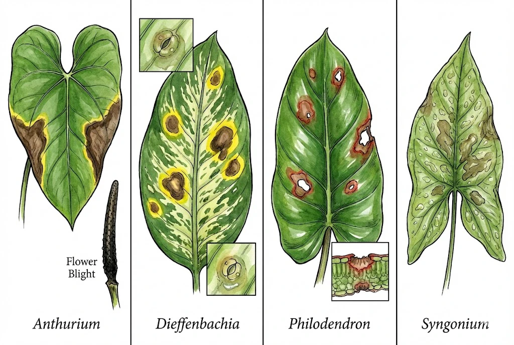
Different genera can react differently to Xpd, and, just as important, different cultivars and bacterial strains do too.
Anthurium
Anthurium is frequently and seriously affected, often with margin-in V-shaped blight and, when systemic, whole-plant decline. The spadix can also be infected. Susceptibility still depends on cultivar and strain.
Dieffenbachia
Dieffenbachia is a documented host, and the pathovar is named for it. Lesions may show a pronounced yellow halo and can be circular where entry is through stomata rather than hydathodes.
Philodendron
A thick cuticle may offer some protection in Philodendron. Heartleaf philodendron has a reported reddish red-edge lesion border, but a rust-border rule for all philodendrons is not well supported.
Syngonium
Thin leaves can make water-soaking obvious, and some reports describe leaf spots that coalesce quickly. This is not a strain-independent rule.
Recent comparative work on Araceae-associated Xanthomonas shows a range of heterogeneous strains, including species such as X. hortorum. A simple split that treats Xpd as primary and everything else as rare oversimplifies the picture.
For a keeper, though, the practical response is consistent. Isolate suspect plants promptly and rely on strict hygiene, clean stock, and (where infection is confirmed) disposal, plus local extension or diagnostic-lab guidance.
Distinguishing Bacterial Blight from Look-Alikes
The table below lists tendencies, not diagnostic certainties. Every row has exceptions, chilling, water molds, mixed infections, and nutrient or salt problems overlap heavily, so use these features to form a hypothesis, then confirm with history, root and crown checks, and, where it matters, a lab test.
| Feature | Bacterial Blight (Xanthomonas) | Fungal Leaf Spot (Colletotrichum) | Abiotic (Fertilizer/Salt Burn) | Abiotic (Iron Deficiency) |
|---|---|---|---|---|
| Emergence Time | 14-30 days post-infection (latency) | 5-7 days post-infection (faster) | 1-2 days (acute) to weeks (chronic) | Slow onset on new growth |
| Specific Texture | Water-soaked, greasy, translucent | Dry, papery, often zonate (rings) | Crispy, dry, brittle burned tips | Soft, limp, or normal texture |
| Specific Color | Brown/Black with Bright Yellow Halo | Tan/Brown with diffuse/dull halo | Brown/Orange crusty edges | Yellow leaf with Green Veins |
| Progression Speed | Rapid expansion (V-shape inward) | Moderate expansion (circular) | Stops if leaching occurs | Persists until nutrients corrected |
| Leaf Location | Margins (hydathodes) or Wounds | Anywhere on leaf blade | Tips and Margins (accumulation) | Newest leaves (immobile nutrient) |
| Systemic Signs | Petiole snap, vascular browning | Rare (unless stem rot) | Root burn, general stunt | Stunting, bleaching of new growth |
Many problems can brown a leaf. Use the patterns below to form a working hypothesis, then check the roots, crown, and recent care history before treating.
Comparative Diagnostics of Aroid Leaf Pathologies
| Feature | Bacterial (Xanthomonas) | Fungal | Abiotic (salt/cold) |
|---|---|---|---|
| Lesion shape | Angular, V-shaped from the margin | Circular, concentric rings | Tip/margin, diffuse patches |
| Halo | Bright lemon-yellow | Diffuse, pale or brown | None, sharp edge |
| Texture | Greasy, water-soaked, translucent | Dry, papery | Crispy and dry |
| Held to light | Translucent, like stained glass | Opaque | Opaque |
| Tell-tale | Bacterial ooze (slime) | Spores or fuzz | Follows a care event |
This table helps you form a first impression, not a final answer. Dry spots with rings lean fungal. Greasy spots with a bright yellow halo lean bacterial. A crispy tip after a recent feed leans abiotic, but each of these can be produced by more than one cause, so treat the read as a hypothesis to confirm. For a species-specific sequence that also checks roots, temperature, light, and pests, use the Monstera black spots diagnostic guide.
Similar Pathogens – Bacterial Soft Rots & Aroid Leaf Disease
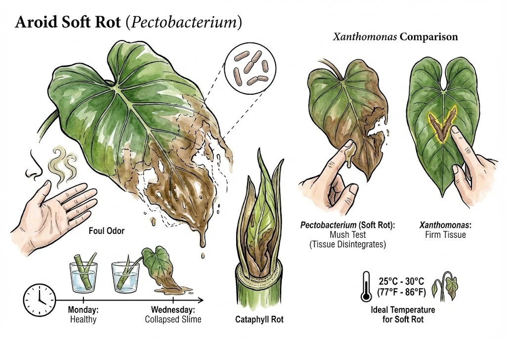
While Xanthomonas often incubates slowly, soft-rot bacteria such as Pectobacterium, formerly called Erwinia, tend to cause faster tissue collapse. Related organisms including Dickeya can look similar, so soft rot is a symptom group rather than a single confirmed species.
Soft rot usually lacks the V-shapes and bright halos of Xanthomonas and instead turns tissue mushy.
Speeds overlap between causes, so use the mushy texture as the main clue rather than the exact timing.
Characteristic Odor
Smell is a supporting clue for soft rot, not a diagnostic test. As tissue breaks down, secondary bacteria growing in the macerated fluid can generate an offensive odour, so the smell reflects the decay process as much as any one pathogen.
That odour is often described as rotting fish or decaying vegetables, and Xanthomonas lesions are frequently less smelly early on. Because odour is subjective and depends on the stage and the mix of microbes present, it cannot confirm which organism is involved.
A foul smell in your plant area is still worth investigating (check nearby plants for soft, rotting tissue) but remember root rot and other decaying material can smell similar.
The Speed of Collapse
Xanthomonas often works over weeks, while soft rot can move in hours to days, though these timelines overlap and are not a fixed diagnostic clock.
Under favorable warm and wet conditions, Pectobacterium can collapse soft tissue very quickly, sometimes within a day or two. Exact times depend on host, isolate, temperature, and bacterial load, so 24–48 hours is an illustration rather than a rule.
Illustrative Soft-Rot Progression
As an illustration of how fast soft rot can move (not a fixed schedule). A Philodendron cutting rooting in water looks healthy one day, has a soft stem the next, and collapses into a mushy, decomposed mass shortly after.
Fast collapse like this is typical of soft rot. The bacteria produce cell-wall-degrading enzymes that macerate tissue, though almost instantly overstates the speed because maceration still takes time and the right conditions.
Symptoms on Unfurling Leaves
Soft rot often affects the cataphyll. The sheath that protects a new leaf. Water trapped in the unfurling coil can create a wet, low-oxygen pocket that favours soft-rot bacteria, and the new leaf may turn to a brown, mushy paste before it opens.
This is a risk factor, not a guarantee that Pectobacterium specifically is present.
Texture Check (The Mush Test)
Gently press the lesion. Ideally with a gloved hand or a clean, disposable tool, then disinfect afterwards so you do not carry bacteria to other plants.
Xanthomonas Lesion Texture
The dead tissue might be dry or slightly gummy, but the surrounding tissue is firm.
Pectobacterium Lesion Texture
The tissue disintegrates under pressure. It feels like wet paper or slime, lacking structure.
The Role of Temperature
Many soft-rot bacteria favor warm, wet conditions, and warm, humid growing rooms can overlap with the range comfortable for tropical aroids. Optimal temperatures differ by species and isolate, so there is no single perfect number, and claims that warming is expanding these diseases need species- and location-specific evidence.
The practical point still holds. A hot, persistently wet environment raises soft-rot risk when a pathogen, a wound, and low oxygen come together, but heat and humidity alone, without the organism present, do not cause the disease.
Fungal Pathogens in Aroid Leaf Disease Diagnostics
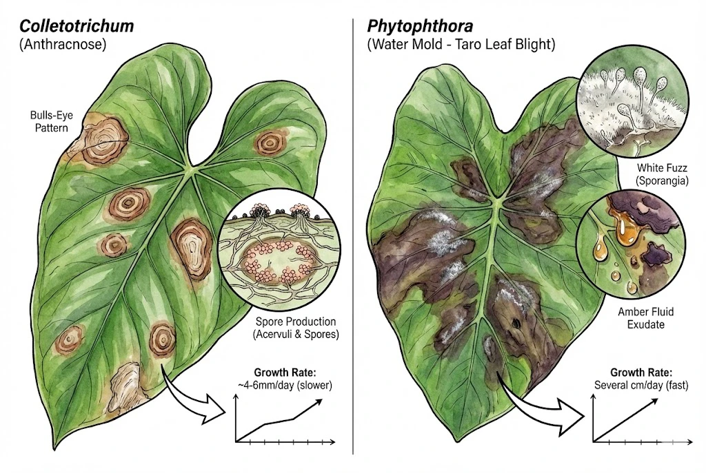
Fungi are significant causes of leaf spots and are often confused with bacteria. However, their biology dictates a different set of symptoms.
Colletotrichum (Circular Lesion Formations)
Colletotrichum species cause anthracnose. Current UF guidance associates C. gloeosporioides on Anthurium mainly with black nose of the spadix, so calling it common leaf anthracnose overstates the evidence. Identifying the species and host matters.
Fungi grow via hyphae (microscopic threads), which can cross veins and often produce more circular spots, in contrast to the angular, vein-limited look many bacterial lesions have.
This is a general tendency with plenty of exceptions, not a hard boundary.
The Concentric Ring Pattern
Some lesions show concentric rings (a target-like zonation), which can arise from environmental cycling as a colony grows.
Rings lean fungal, but they are neither required for a fungus nor impossible for other causes, so they are a clue rather than proof.
Spore Production
In high humidity, fungi produce fruiting bodies.
If you look closely at an Anthracnose spot (using a magnifying glass), you might see tiny black dots (acervuli) or pinkish/orange masses of spores arranged in rings.
Bacteria do not produce these structures. They produce amorphous ooze.
Growth Rate
Do not diagnose from how quickly a spot grows. Lesion speed varies with the cause, the plant, and the conditions.
Fungal lesions are often more contained than a bacterial systemic collapse, but vascular fungi and oomycetes can also spread aggressively. Fungal disease is not always slower.
The edge of a fungal lesion is frequently dry and distinct, without the broad greasy yellow halo that leans bacterial (again a tendency, not a pathogen-specific rule).
Phytophthora (The Water Mold)
Phytophthora is an oomycete (water mold), not a true fungus. Phytophthora colocasiae causes Taro Leaf Blight and can devastate a taro (Colocasia) crop, but its host range is limited. It is not a general differential for every indoor aroid.
Phytophthora Symptom Clues
| Feature (clue, not confirmation) | Leans fungal (Colletotrichum) | Leans bacterial (Xanthomonas) |
|---|---|---|
| Lesion Shape | Often circular / irregular | Often angular / V-shaped / vein-limited |
| Internal Pattern | May show concentric rings (zonate) | Usually no rings |
| Surface Signs | Black dots (acervuli) or fuzzy mold | Shiny slime (ooze) or nothing |
| Halo | Often dull, diffuse, or absent | Often bright yellow |
| Smell | Sometimes earthy / mushroomy | Often odorless early, sour later |
| Under magnification | Hyphae and spores may be visible | Bacterial streaming possible, not species confirmation |
Phytophthora lesions on taro start small, dark, and water-soaked (similar to bacterial spots).
On susceptible, wet taro they can expand fast, and a white fuzz (sporangia) may appear on the lesion surface, especially in morning dew.
Exact speed and visibility depend on conditions, and the absence of fuzz does not rule Phytophthora out.
A rough memory aid is that Xanthomonas tends to produce slime while Phytophthora tends to produce fuzz, but neither sign is always present.
Phytophthora lesions can also exude a clear, amber-to-darker fluid, whose colour shifts with age and conditions. This overlaps with, rather than cleanly separating from, bacterial ooze.
Fungal vs. Bacterial clues that require confirmation
These are tendencies with important exceptions. Fungi and oomycetes can cross veins and cause vascular or systemic disease, a bacterial leaf infection may never go systemic, and a negative ooze test does not rule bacteria out.
| Marker | Often bacterial | Often fungal / water mold |
|---|---|---|
| Surface growth | Amorphous ooze and slime | Spore masses, rings, or white fuzz (Phytophthora) |
| Lesion edges | More often vein-limited (angular) | More often cross veins (circular) |
| Spread | Can move systemically through the xylem | Often within leaf tissue (but some are vascular) |
| Ooze test | Milky stream possible from a cut vein | Usually no stream (not a reliable exclusion) |
Abiotic Errors vs. Aroid Leaf Disease
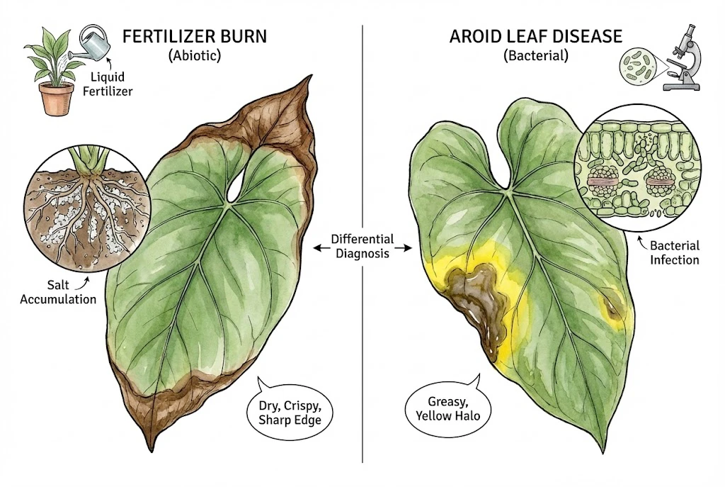
Alongside checking for disease, rule out abiotic (non-living) causes such as care errors. Isolate a genuinely suspect plant first. You can rule out abiotic factors while it is already separated.
Abiotic damage often has helpful patterns (for example, tip-and-margin burn from salts), but it does not have patterns that living pathogens can never mimic. Bacterial, fungal, chilling, and nutrient problems overlap in appearance more than diagnostic tables suggest.
Fertilizer Burn (Salt Accumulation)
Rapid growth goals often lead to heavy fertilizer use. This leads to a buildup of soluble salts in the soil.
How Salt Injury Blocks Water Uptake
High salt concentration in the soil creates osmotic pressure that prevents roots from taking up water (physiological drought).
The salts also accumulate in the leaf tips and margins, where the water evaporates, leaving the salt behind to burn the tissue.
How Salt Burn Appears
Crispy Tips
Salt-related damage often shows at the tips or margins, though root injury can produce other patterns and symptoms vary by species.
Dry, Brittle Texture
The dead tissue is usually dry, brittle, and brown rather than mushy, but secondary rot can change this.
Sharp Demarcation
The line between dead and living tissue is often sharp, with little or no yellow halo. This is a clue, not a certainty.
Timing After Feeding
Symptoms may follow a heavy feed within a day or two, or appear gradually with slow-release fertiliser. Exact timing is not fixed.
Check EC and Roots Before Flushing
A greasy lesion with a bright yellow halo leans bacterial, while a dry, crispy tip after feeding leans abiotic, but neither look confirms the cause. Don’t flush purely on appearance.
Check the runoff or substrate EC, your water quality, and root health first. Flushing an already saturated, low-oxygen mix can make root problems worse.
Cold Damage (Chilling Injury)
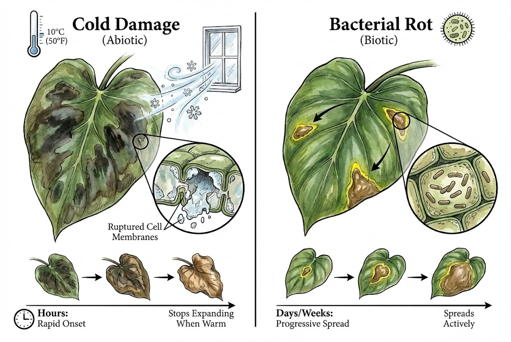
Aroids are tropical and can be harmed by cold. Chilling exposure, cool but above freezing, during shipping or by a drafty window can cause damage even without frost.
How Chilling Damages Leaf Tissue
At around 10°C the water in a plant is not freezing (that is well above the freezing point), so this is chilling injury, not ice damage.
Above-freezing chilling mainly disrupts cell membranes and metabolism. Actual freezing, which does form ice and rupture cells, is a separate, colder event.
How Chilling Injury Appears
Delayed Symptom Onset
Symptoms may take from hours to several days to appear. In some plants damage continues to show up over a week or more, so an onset within hours is not universal.
Exposure Pattern
Chilling injury often produces large, diffuse, dark, water-soaked patches, frequently on the side nearest the cold source, such as a window. This is a useful clue, not a diagnosis.
Tissue Changes
Tissue may darken and soften, then dry to crispy brown, with the exact course depending on species and severity.
Damage Can Continue After Warming
Chilling injury does not multiply like a pathogen, but damage does not stop the moment the plant warms up. Primary injury and secondary decay can continue to reveal themselves for days.
Iron Deficiency vs. Viral Chlorosis
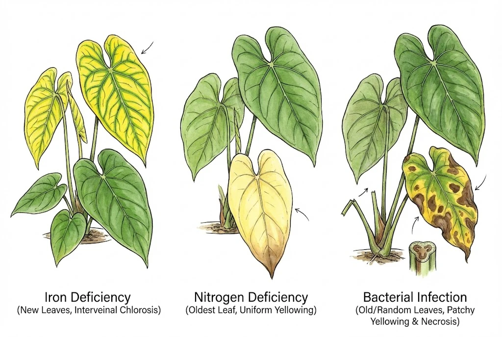
Yellowing has many causes, so leaf location is one clue among several, not a decisive test. Confirm with root, pH, and, where needed, tissue or lab checks.
Iron Deficiency
Iron moves poorly within the plant, so its deficiency tends to show on the newest leaves first.
Iron Deficiency Symptom
Interveinal chlorosis produces a yellow blade with veins staying green. Manganese deficiency looks very similar, and pH and root oxygen affect uptake, so iron is not confirmed by appearance alone.
Iron Deficiency Differentiation
Yellowing from a systemic infection more often affects older or scattered leaves. Vein colour is only a hint. Green veins lean toward a nutrient issue, but this is not a hard rule, and browning veins can occur for several reasons.
Viral Chlorosis
Viruses can also yellow leaves, but the pattern usually differs from a simple nutrient deficiency. Think mosaic mottling, streaking, ringspots, or one-sided (sectored) chlorosis that does not correct with feeding.
Viruses cannot be confirmed by eye (diagnosis needs testing). If you suspect a virus, isolate the plant, avoid propagating from it, and keep tools clean, since many viruses spread on cutting blades and by handling.
Nitrogen Deficiency
Nitrogen is mobile. The plant steals it from old leaves to fuel new growth.
Nitrogen Deficiency Symptom
The oldest leaves fade to a fairly uniform pale yellow. Similar old-leaf fading can come from other mobile-nutrient shortfalls, light, or root problems, so confirm with soil or tissue testing rather than colour alone.
Nitrogen Deficiency Differentiation
Infection-related yellowing is often patchy and may come with wilting or weak petioles, while nitrogen deficiency is usually a slow, even fade of the oldest foliage. These are tendencies, not a substitute for a lab or soil diagnosis.
Identifying and correcting iron deficiencies in ornamentals (Oklahoma State Extension)
A Home Triage Toolkit (Not a Confirmed Diagnosis)
You can do useful home triage with common items, but be clear about what it can and cannot do.
These checks can raise or lower your suspicion. They cannot confirm the pathogen or its pathovar.
Confirming Xpd (and distinguishing it from Ralstonia, Pectobacterium, Dickeya, fungi, or oomycetes) requires culturing, immunoassays, or PCR done by a diagnostic lab.
If you suspect a serious bacterial disease, isolate the plant, photograph and record the symptoms and conditions, and contact your local extension service or a plant-disease clinic. Some diseases also carry regulatory reporting requirements.
At the first suspicious spot, I photograph both leaf surfaces, the petiole, roots, and the plant’s position before moving it to isolation. I place a small paper scale beside the lesion and repeat the same view daily, which shows whether the margin is expanding or whether an old injury has simply darkened.
I also record watering, spraying, repotting, cold exposure, and nearby affected plants on the same timeline. That history is often more useful to a laboratory or Extension diagnostician than a cropped picture of the final brown patch.
The Streaming (Ooze) Observation
Streaming can suggest that bacteria are present, but it is not an Xpd-specific test and cannot separate bacterial species.
Handle the sample with gloves and clean, disposable tools, and disinfect afterwards so you do not spread anything.
1. Prepare the Sample
Using a clear glass of clean water, cut a small piece of tissue, about 1 cm, that includes the border between dead and living tissue, cutting through a vein if possible.
2. Suspend the Tissue
Suspend the piece so it just touches the water. A clean paperclip or tweezers can help.
3. Observe the Water
Shine a light through the glass from the side or back.
Milky Streaming Is Suggestive but Nonspecific
A milky thread drifting from the cut vein over a few minutes suggests a high bacterial load.
It does not tell you which bacterium, or whether it is pathogenic (that still needs a lab).
No Streaming Does Not Rule Out Bacteria
Low bacterial numbers or a local, non-vascular infection can give no visible stream, and fungal or abiotic causes remain possible.
Handling Weak Petioles (a Cautionary Note)
Easy break-off of leaves is a recognised sign of systemic Anthurium blight, but deliberately snapping a petiole is destructive and not a reliable confirmation. Bending a petiole can injure healthy tissue and fling infectious ooze, while mechanical strength varies with age, hydration, species, and ordinary rot. Note unusual weakness during normal handling, wear gloves, disinfect tools, and rely on lab testing.
Smell as a Weak Clue
Smell is subjective and depends on the stage of decay and the mix of microbes present, so it is at best a weak clue.
Fungal Rot Odor
Fungal rots can smell mushroomy or earthy.
Bacterial Soft-Rot Odor
Bacterial soft rots caused by Pectobacterium often smell of rotting fish or old potatoes, partly from secondary bacteria in the decaying fluid.
Xanthomonas Odor
Xanthomonas lesions are frequently less smelly early on.
Limitations of Chemical Control
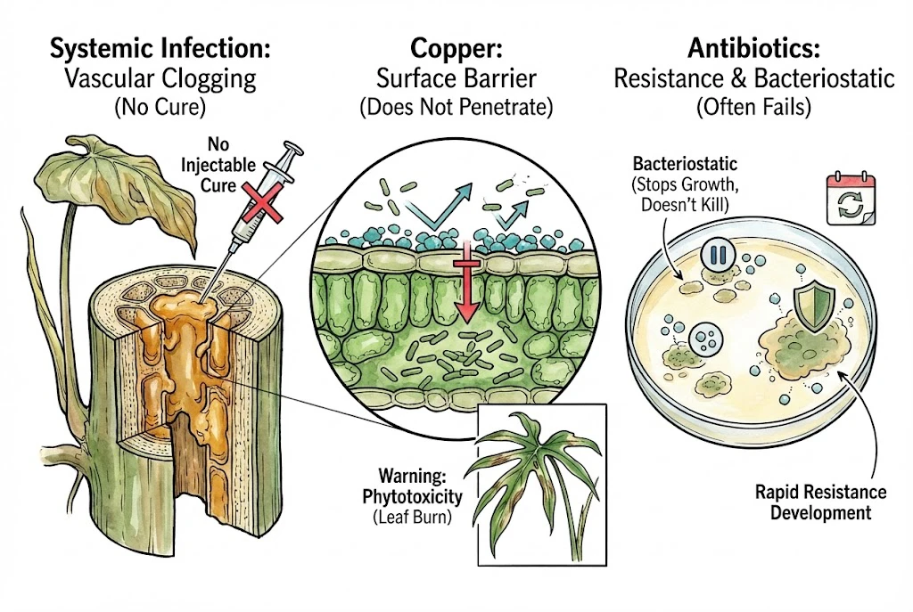
Confirmed systemic bacterial blight in Anthurium has a poor prognosis. Once bacteria are established in the vascular system, no home treatment reliably clears them. Injecting antibiotics is not a practical or recommended fix, and online cures promising to reverse systemic infection deserve skepticism. The first response to a confirmed case is isolation and usually disposal of the infected plant, plus advice from a diagnostic lab or extension service about regulatory steps.
Copper Fungicides/Bactericides
Copper is one option among several. UF/IFAS also lists mancozeb and Bacillus subtilis products, so copper is not the only treatment. It works mainly as a protective surface layer, where copper ions can disrupt bacterial and fungal cells on contact.
Because it is a surface treatment, copper does not penetrate leaf tissue well. It cannot reach bacteria already inside the mesophyll and is best thought of as helping protect still-healthy tissue, not curing infected leaves.
Critically, copper is a regulated pesticide. Whether you may legally use a given product on aroids for a bacterial disease depends on the exact product label, your crop/site, the disease, and your jurisdiction.
Before use, confirm the label allows it, follow the stated dilution and PPE, and heed cautions on phytotoxicity, temperature/sun, and runoff to waterways.
Copper Products
Ready-to-use copper products are widely available, but check the current label before using any of them against Xanthomonas on Anthurium, since permitted crops and diseases vary from product to product.
Copper can also burn thin aroid foliage, so read the label, spot-test first, keep it off hot or sunny plants, and treat it as possible prevention rather than a cure. Don’t apply it on an unconfirmed diagnosis.
Copper Phytotoxicity Warning
Copper can cause leaf burn (phytotoxicity), especially on thin-leaved plants. Test a small area first and avoid applying in hot, sunny conditions.
Antibiotics
Agricultural antibiotics (for example, streptomycin-based products) exist, but their availability and legal use depend on the current product label, crop, disease, and region, and they are generally not appropriate for home use.
Resistance can develop and they are costly.
It is not accurate to say all antibiotics are merely bacteriostatic at safe doses. Streptomycin, for instance, is classified in regulatory records as a bactericide or bacteriostat depending on use. Effectiveness and legality must be judged from the current label and resistance-management guidance.
Cultural Control (The Core of Prevention)
Because reliable cures are limited, prevention matters most. Extension guidance points to a familiar core of practices for aroid keepers.
1. Keep Foliage Dry
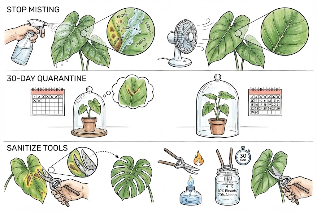
When a pathogen is present, a water film lets bacteria travel from a sick leaf to a healthy one, so keeping leaves drier and air moving lowers risk. A humidity spike can encourage guttation, but misting does not mechanically open the hydathodes. A fan can help, but it is not automatically better than every other measure.
A small circulation fan helps keep leaf surfaces dry and the air moving, which supports bacterial-spread prevention alongside, not instead of, clean stock, isolation, and sanitation.
Circulation Fan
A quiet, speed-controlled circulation fan can help leaves dry, but a catalog airflow figure does not tell you the velocity at the canopy. Size and aim the fan by measured air movement and dead zones rather than by one product’s CFM rating.
Check the exact fan’s operating-humidity and water-resistance limits. Electronics-cooling fans are often not rated for a misted or condensing cabinet, so keep an unrated unit in a splash-free, dry position and protect every electrical connection.
Drying the leaf surface removes a condition bacteria use to move, but no fan is a proven stand-alone preventive.
1. Quarantine New Plants
Because Xanthomonas can stay symptom-free well beyond a week, sometimes for months, isolate new arrivals and watch them for at least 30 days. Treat 30 days as one observation window, not a guarantee. Clean, screened stock and, where possible, testing do more than any waiting period, and staying symptom-free reduces risk without proving the plant is disease-free.
2. Disinfect Tools Between Plants
Cutting an infected leaf and then a healthy plant with the same blade can transfer bacteria in the sap. Between plants, clean off debris and disinfect blades. If you use a bleach solution, a common recommendation is roughly a 1/9 household-bleach dilution. If you use isopropyl alcohol at 70% or more, remember it is flammable. Follow the product’s stated contact time, work in a ventilated area, rinse and dry metal to limit corrosion, and never mix bleach and alcohol. Keep flammable alcohol away from any open flame.
Managing Aroid Leaf Disease
When a spot is serious or spreading, isolate the plant, photograph the progression, check roots and recent conditions, and seek a diagnostic lab or extension service before applying chemicals or discarding it.
Dry foliage, quarantine, clean tools, and a confirmed diagnosis prevent more damage than any single spray.